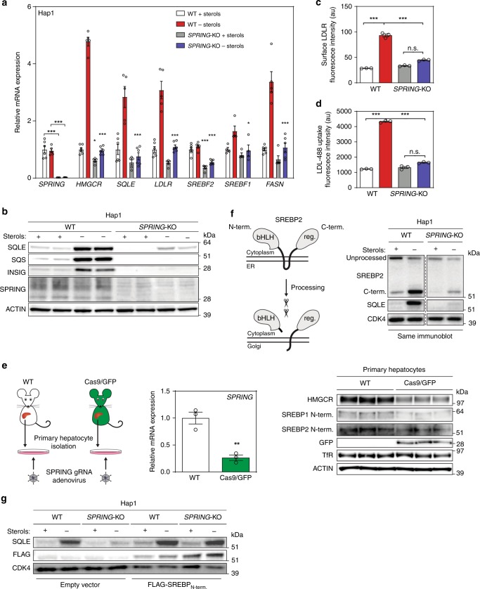
Fig. 2. Ablation of SPRING reduces SREBP signaling in cell lines and primary mouse hepatocytes.
a, b Hap1-WT and Hap1-SPRINGKO cells were cultured in the presence or absence of sterols. Subsequently, cells were harvested for a gene expression analysis by qPCR as indicated (N = 5 biologically independent samples), and b immunoblotting as indicated (N = 3). c, d Cells were grown as in a, b and were (c) stained with an anti-LDLR-APC antibody, or d incubated with 5 µg/ml DyLight 488-labeled LDL for 1 h. Subsequently, cells were fixed and analyzed by FACS (N = 3 biologically independent samples). e Primary mouse hepatocytes were isolated from C57BL/6J WT and Cas9 knock-in mice and 4 h post isolation infected with Ad-3x -sgRNA-Spring adenoviral particles. Cells were sterol-depleted for 16 h and harvested for immunoblotting and gene expression analysis as indicated (N = 3 biologically independent samples). f Schematic representation of SREBP processing (left). Total cell lysates from Hap1-WT and Hap1-SPRINGKO cells were immunoblotted as indicated (right). A representative image of at least three independent experiments is shown. g Hap1-WT and Hap1-SPRINGKO cells were transduced with a FLAG-tagged constitutively active SREBP2 N-terminal construct. Total cell lysates were immunoblotted as indicated (N = 2). All bars and errors represent mean ± SEM; *p < 0.05, **p < 0.01, ***p < 0.001.

