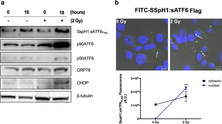
Figure 4.
Endoplasmic reticulum stress (ERS)-related protein expression in CT26 cells infected with KST0652. The CT26 cell monolayer was infected with KST0652 (MOI = 100) and irradiated with 2 Gy of γ-radiation at 2 h post-infection. (a) The expression levels of SspH1:sATF6FLAG, p90ATF6, p50ATF6, GRP78, and CHOP proteins were detected using western blot analysis. β-tubulin was used as the loading control. Full-length blots are presented in Supplementary information. (b) SspH1:sATF6FLAG was detected in KST0652-infected CT26 cells after 18 h post-irradiation. Immunofluorescence assay, followed by confocal microscopy, was used to visualize the location and expression of SspH1:sATF6FLAG in CT26 cells. The nuclei of CT26 cells were stained with DAPI (blue), and SspH1:sATF6FLAG were stained with FITC-conjugated anti-FLAG mouse IgG (green). White arrows indicate SspH1:sATF6FLAG protein. Representative images are shown; original magnification, 100×. Changes in fluorescence signals of SspH1:sATF6FLAG in CT26 cells. Data are mean fluorescence intensities (a.u.) ± SEM from three independent photos and asterisks (*) indicate significant difference between each group (** P < 0.01).

