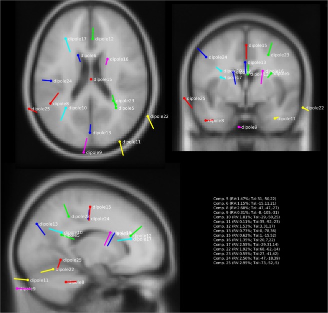
Figure 5.
Source localizations of the fifteen components in the experiment using equivalent current dipole source fitting. Plotted are the fifteen components with clear dipolar topographic maps (see Figs. 2–4). For each component we also list the Residual Variance (RV) between the model prediction and the observed data, as well as the anatomical coordinates in Talairach space (between brackets). Note how sources are localized in many different areas of the brain, including occipital, parietal, temporal and frontal areas.

