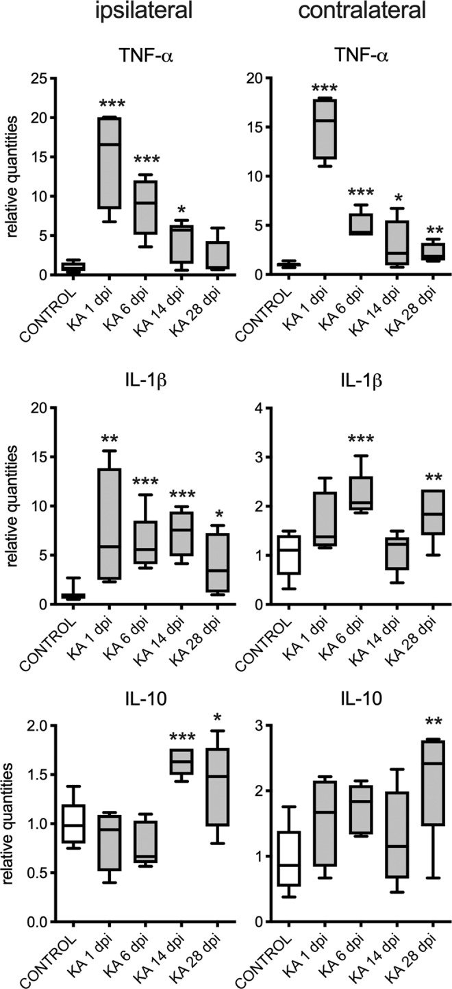
Figure 6.

The relative expression of proinflammatory (TNF-α and IL-1β) and anti-inflammatory cytokines (IL-10) in ipsilateral and contralateral hippocampi at 1, 6, 14 and 28 days post injection (dpi) of KA in adult male mice (n = 5 in each group). The left panel shows changes in ipsilateral hippocampus. The right panel shows changes in contralateral hippocampus. The t-test with multiple-testing correction was performed to evaluate significance between control and KA groups *p < 0.05, **p < 0.005, ***p < 0.001. Figures were created using GraphPad Prism version 8.3.1 for macOS, www.graphpad.com.
