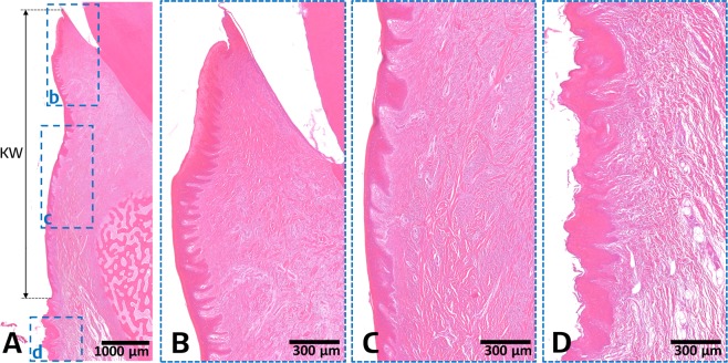
Figure 2.
Representative histomicrographs of a buccolingual section at the experimental site: overview (A), gingival sulcus region (B), attached gingiva region (C), and mucosa region (D). The gingival sulcus and attached gingiva regions appear as keratinized, stratified squamous epithelial cell layers with numerous rete pegs and underlying dense connective tissue layer. In contrast, the mucosa region shows a nonkeratinized, thin epithelial layer and underlying loose connective tissue. Based on these specific histologic features, the mucogingival junction can be clearly distinguished and the keratinized tissue width (KW) can be measured.

