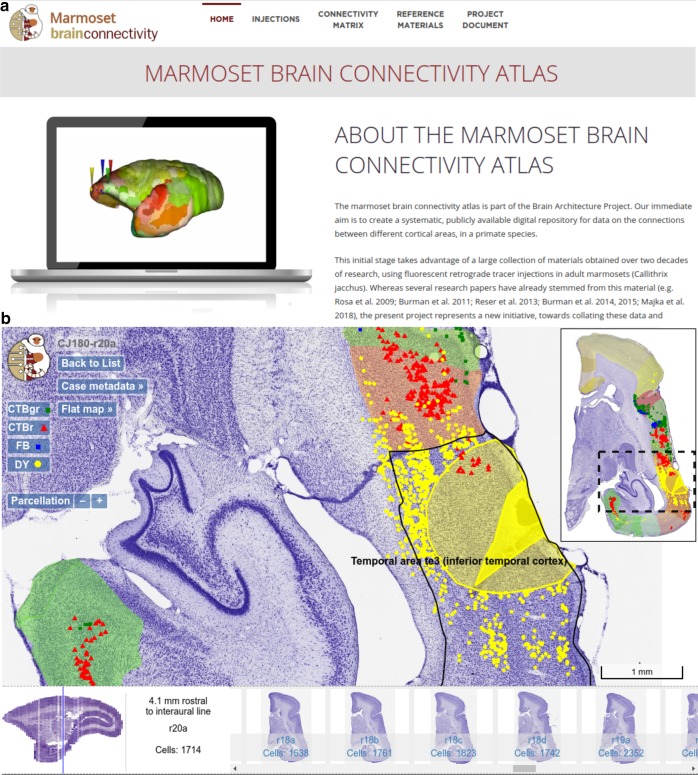
Fig. 2. Overview of the Marmoset Brain Connectivity Atlas and high-resolution section viewer.
a Gateway page of the portal (http://marmosetbrain.org) offering access to different components of the website, including the primary experimental data (Injections tab) and quantitative results, such as the FLNe, SLN, and interareal distance matrices (Connectivity Matrix tab, see Fig. 3). The Reference Materials tab gives access to the reference atlas29, the volumetric template of the marmoset brain, and histology protocols. The Project Document tab provides background information on the aims of the project and its implementation. b Highlight of the main features of the high-resolution section viewer (injection CJ180-DY used as an example). An image of a Nissl-stained section (r20) is overlaid with the injection site and halo (yellow polygons, drawn under the microscope), as well as locations of cells labeled by DY and other tracers injected in this case (points in various colors). The navigation bar (bottom) offers a quick way of traversing across the dataset while the widgets (buttons on the top left) allow for adjusting the view according to one's requirements. The cortical areas on the section are annotated based on the registration to the reference atlas29 (thumbnail in the top right corner). The contents of the portal are available under an open license (https://creativecommons.org/licenses/by-sa/4.0/).

