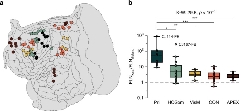
Fig. 7. Local versus distant connectivity in cortical networks.
a Flat reconstruction of the marmoset cortex, showing the location of injection sites attributed to five networks of areas, defined in previous studies45,47,48. Pri (dark green): primary sensorimotor network (areas 3a, 3b, 4ab, and 4c); HOSom (light green): a network formed by interconnected higher-order somatosensory and premotor areas (dorsocaudal premotor area [6DC], medial premotor area [6M], caudal ventral premotor area [6Va], dorsal cingulate motor area [24d], and parietal area PF); VisM (yellow): a network formed by higher-order visuomotor integration areas (lateral and medial intraparietal areas [LIP and VIP], caudal subdivision of PE [PEc], and putative subdivisions of the frontal eye field [8aV and 8C]); CON (orange): cognitive control network (putative homolog of the default mode network B, including frontal areas 8aD and 6DR, cingulate areas PGM and 23b, and ventral parietal areas OPt and PG); APEX (brown): apex transmodal network (putative homolog of default mode network A, including areas 10, 23a, TPO, PGa/IPa, and the rostral part of TE3, near the temporal pole). b Box plot (center line: median; box limits: upper and lower quartiles; whiskers: 1.5× interquartile range; annotated points: outliers) of the ratio of the fraction of labeled neurons located within 4 mm of the center of the injection site (local connections) to neurons located >8 mm from the site (distant connections) following injections in the five networks (K–W: Kruskal–Wallis H test: 29.8, p < 10−5). Statistical significance according to post-hoc Dunn's test: p ≤ 0.05 (*), p ≤ 10−2 (**), p ≤ 10−3 (***). Source data are provided as a Source Data file.

