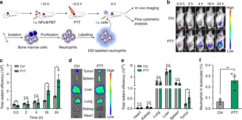
Fig. 3. PTT-created tumoral inflammatory microenvironment recruited adoptively transferred neutrophils.
a Schematic showing analysis of the accumulation of transferred DiD-labeled neutrophils in PTT-treated EMT6 tumors. EMT6-bearing mice were i.v. injected with NPs@PBT at −12 h and tumors were irradiated with an 808 nm laser (40 °C for 5 min) at −0.5 h. Neutrophils were isolated from bone marrow, labeled with DiD, and i.v. injected into the mice on 0 h. b, c The distribution of transferred DiD+ neutrophils in the recipient mice was analyzed with the IVIS at the indicated time points. b Representative images and c corresponding quantification of tumoral fluorescence intensities at the indicated time points. n = 3 mice per group. d Ex vivo fluorescence images and e corresponding quantification of total fluorescence intensities of tumors and major organs collected at 24 h post-cell transfer. n = 4 for control group and n = 3 for PTT group. f The percentage of transferred DiD+ neutrophils in total DAPI−CD45+ tumor-infiltrating leukocytes analyzed by flow cytometry at 10 h post injection. n = 3 for control group and n = 4 for PTT group. Data are shown as mean ± SEM and analyzed by unpaired two-tailed Student’s t-test. *P < 0.05, **P < 0.01. n. s., not significant. Source data are provided as a Source Data file.

