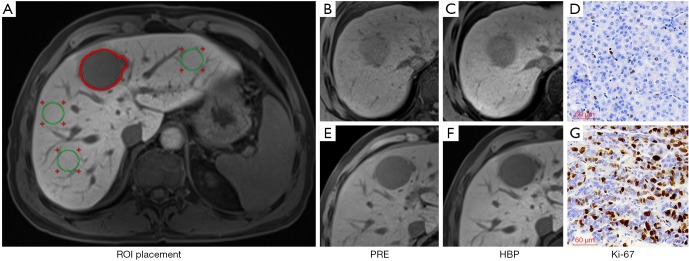
Figure 2.
Illustration of ROI placement (A) with three circles ROIs (green ROIs) on the left lobe, right lobe anterior and posterior segments, respectively, and a freehand ROI along the tumor margin at the level of the largest diameter of the tumor (red ROI). And the representative images of PRE, HBP, and Ki-67 staining (magnification, ×200) in low aggressive HCC (B,C,D) and high aggressive HCC (E,F,G). The RTE, TLR, TEI and RER were 0.32, 0.76, 0.93 and 0.62 in a 59-year-old male patient with low aggressive HCC (Ki-67 LI =3%) and 0.21, 0.50, 0.62 and 0.18 in a 50-year-old male patient with high aggressive HCC (Ki-67 LI =57.62%). ROI, region of interest; HCC, hepatocellular carcinoma; PRE, pre-enhancement; HBP, hepatobiliary phase; RTE, relative tumor enhancement; TLR, tumor to liver contrast ratio; TEI, tumor enhancement index; RER, relative enhancement ratio.

