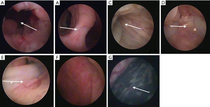
Figure 2.
Representative hysteroscopy in the goat and the novel stent studied inside the uterine cavity of goats. (A) Cervical canal; (B) lower edge of the bifurcation between the left and right horns; (C,D,E) hysteroscopic images showing stent inside the uterine cavity of goat, include the right uterine cavity, the left uterine cavity and the bottom of the uterus; (F) hysteroscopic picture after the stent was removed; (G) hysteroscopic pregnancy sac.

