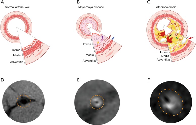
Figure 2.
(A,D) Showed the simplified image and the actual HRMRI image of the normal artery wall. (B) represented the affected artery wall in MMD, which characterized by the thickness of intima and the atrophy of media (the yellow triangle indicated the shifted and proliferated smooth muscle cells, the purple triangle indicated the fibroblast hyperplasia, the red arrow indicated the extremely tortuosed internal elastic membrane, the blue arrow indicated the decreasing of smooth muscle cells in the shrinking media). (C) Was the simplified pattern image of atherosclerosis artery wall which included the lipid deposition under the endothelium, the foam cell formation (green triangle), smooth muscle cells shifting and proliferation (yellow triangle), internal elastic membrane rupture (red arrow), calcium core (gray arrow) and internal bleeding (black arrow). (E,F) Showed the actual HRMRI image of patients with MMD or atherosclerosis. HRMRI, high-resolution magnetic resonance imaging; MMD, moyamoya disease.

