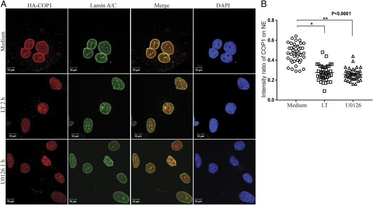
Fig. 3.
Anthrax LT and U0126 treatment promotes a rapid relocation of COP1 from the nuclear envelope (NE) to nucleoplasm. (A) Hepa1c1c7 cells stably expressing HA-COP1 were cultured with or without LT for 2 h or U0126 for 1 h. The cells were then used for immunofluorescence staining with mouse anti-Lamin A/C and rabbit anti-HA primary antibodies followed by goat anti-mouse IgG-Alexa Fluor 633 and goat anti-mouse IgG-Alexa Fluor 568 secondary antibodies. The locations of Lamin A/C, HA-COP1, and the nucleus were assessed by LSM 880 confocal microscope. (B) Fluorescence intensities were quantified using the Bitplane Imaris image analysis software. The intensity ratios of COP1 located on the NE to that in the nucleoplasm were calculated using data from 40 nuclei, from 9 to 13 randomly selected image files obtained from two or three independently performed experiments. Data were statistically analyzed using an unpaired, two-tailed t test at the 95% confidence interval using GraphPad Prism software and presented as means ± SE. P < 0.05 was considered statistically significant.

