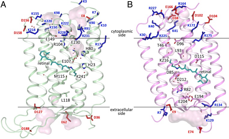
Fig. 2.
Comparison of 48C12 (green) and bR (purple; PDB ID code 1C3W). (A) Side view of the HeR-48C12. N terminus is at the cytoplasmic side of the membrane. (B) Side view of the bR. N terminus is at the extracellular side of the membrane. Hydrophobic/hydrophilic membrane boundaries are shown with black lines. Positively and negatively charged residues on the protein cytoplasmic and extracellular surfaces are shown in blue and red, respectively.

