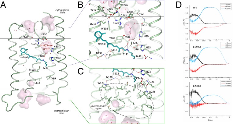
Fig. 3.
Structure of the 48C12 protomer. (A) Side view of the protomer in the membrane. (B) Detailed view of the cytoplasmic part. (C) Detailed view of the extracellular side and the hydrophobic region. Cofactor retinal is colored teal. Hydrophobic/hydrophilic membrane boundaries are shown with gray lines. Cavities are calculated with HOLLOW (31) and shown in pink. Charged residues in 48C12 are shown with thicker sticks. Helices F and G are not shown. (D) Time evolution of the transient absorption changes of photo-excited 48C12, wild-type (WT), E230Q, and E149Q mutant forms. The characteristic wavelengths of intermediate states are slightly shifted in the mutants. The O2-state decay is almost two times longer in both 48C12 variants. Abs, absorption; AU, absorption unit.

