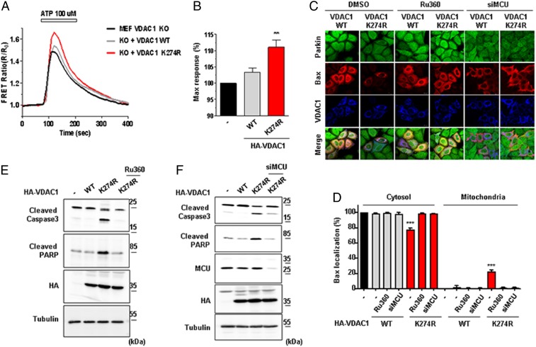
Fig. 3.
Monoubiquitination-deficient VDAC1 induces apoptosis by increasing mitochondrial calcium uptake. (A and B) Mitochondrial calcium dynamics in VDAC1 KO MEF cells. VDAC1 KO MEF cells were transfected with HA-tagged VDAC1 WT or K274R and cotransfected with 4mitD3, a mitochondrial calcium indicator, for 48 h. Cells were then treated with 100 µM ATP for 3 min and were monitored for the changes in 4mitD3 fluorescence. (B) Maximum mitochondrial calcium uptake for VDAC1 KO MEF cells expressing VDAC1 WT or K274R in A. All of the values were normalized to the basal level of VDAC1 KO MEF cells transfected with empty vector (−) (n = 50 to ∼70 cells). Data were analyzed by ANOVA Tukey test. **P < 0.05. (C and D) Confocal images of Bax translocated to the mitochondria. (C) Flag-tagged Bax (red) and HA-tagged VDAC1 WT or K274R (blue) were transfected and immune-stained with Flag and HA antibodies in HeLa cells stably expressing GFP-Parkin (green). Cells were treated with or without 0.5 mM Ru360 for 8 h or transfected with MCU siRNA. (Scale bars, 20 µm.) (D) Percentage of cells with Bax localized in the cytosol or the mitochondria shown in C (n > 500 cells from three independent experiments). Data were analyzed by one-way ANOVA with Tukey multiple-comparison test and are presented as means ± SD. ***P < 0.001. (E and F) HA-tagged VDAC1 WT or K274R was transfected in HeLa cells stably expressing GFP-Parkin. Cells were treated with or without 0.5 mM Ru360 for 8 h in E or transfected with MCU siRNA in F. Cell lysates were analyzed by immunoblotting to examine indicated proteins.

