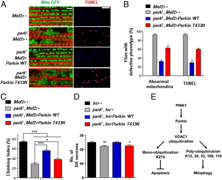
Fig. 6.
Drosophila Parkin T433N cannot rescue the PD-related phenotypes of the park1 mutant. (A) Mitochondrial morphology and TUNEL signals in the thoraces of Parkin-null flies (park1) expressing Parkin WT or T433N using the Mef2-Gal4 driver. Mitochondrial morphology (green) and phalloidin staining (red) to indicate muscle fiber are shown (Left). TUNEL signals (red) and 4′,6-diamidino-2-phenylindole staining (blue) are shown (Right). (Scale bars, 5 μm.) (B) Percentage of flies with defective mitochondrial morphology or TUNEL signals (n = 100 from 10 independent experiments). (C) Percentage of flies with defective climbing ability (n = 100 from 10 independent experiments). (D) Numbers of DA neurons in the PPL1 region (n = 10). All quantifications were analyzed by one-way ANOVA with Tukey multiple-comparison test and are presented as means ± SD. *P < 0.05; **P < 0.01; ***P < 0.001. (E) A schematic model of mitophagy and apoptosis regulated by two types of ubiquitination on VDAC1 in the PINK1–Parkin pathway.

