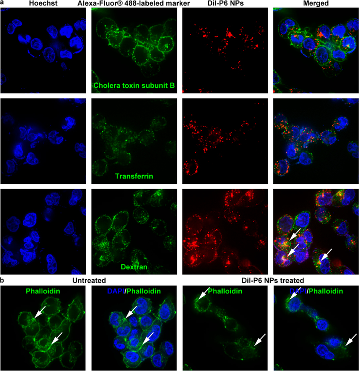
Figure 4.
Confocal laser scanning microscopy utilization to visualize cytoplasmic delivery of P6 NPs. (a) Dil-P6 NPs were incubated with A2780cis cells in the presence of Alexa-Fluor 488-labeled markers of various endocytic pathways. NPs co-localized with dextran, a fluid-phase marker known to enter cells via macropinocytosis (white arrows). (b) Alexa-Fluor 488-labeled actin fibers revealed membrane ruffling and actin rearrangement (white arrows), hallmarks of uptake by macropinocytosis.

