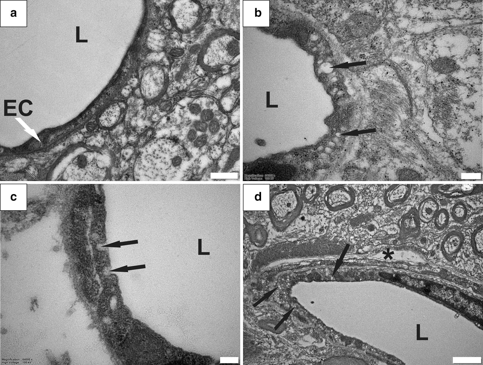
Fig. 4.

Abundant pinocytotic vesicles in endothelial cells in PTS. Blood vessels in healthy spinal cord tissue show a limited number of intracellular vesicles (a). In tissue from PTS animals, some blood vessels contained abundant electron-lucent vesicles indicated by black arrows (b–d). Intracellular vesicles fusing with the endothelial plasma membrane (c). Note the blood vessel in (d) also shows a microcavity in the perivascular region, suggesting that the two processes may be related. EC, endothelial cell; L, lumen; *, perivascular microcavity. Magnification: ×19,000 (a), ×34,000 (b), ×64,000 (c), ×10,500 (d). Scale bars: 0.5 µm (a), 0.2 µm (b), 0.1 µm (c), 1 µm (d)
