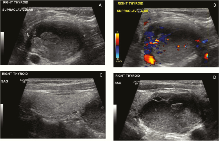
Figure 1.
Ultrasound images demonstrating a large 2.9 × 3.9 × 4.7 cm complex mass at the lower pole of the right thyroid in the supraclavicular area with mixed solid and cystic components that proves to be the parathyroid adenoma. The mass on sagittal images appears to be separate from the right thyroid lobe and located posterior to the lower pole. A: Transverse image, right lower neck. B: Transverse image right lower neck with Doppler. C: Sagittal image demonstrating a normal right thyroid lobe with the complex mass at the inferior border of the thyroid. D: Sagittal image of the complex mass.

