Figure 7.
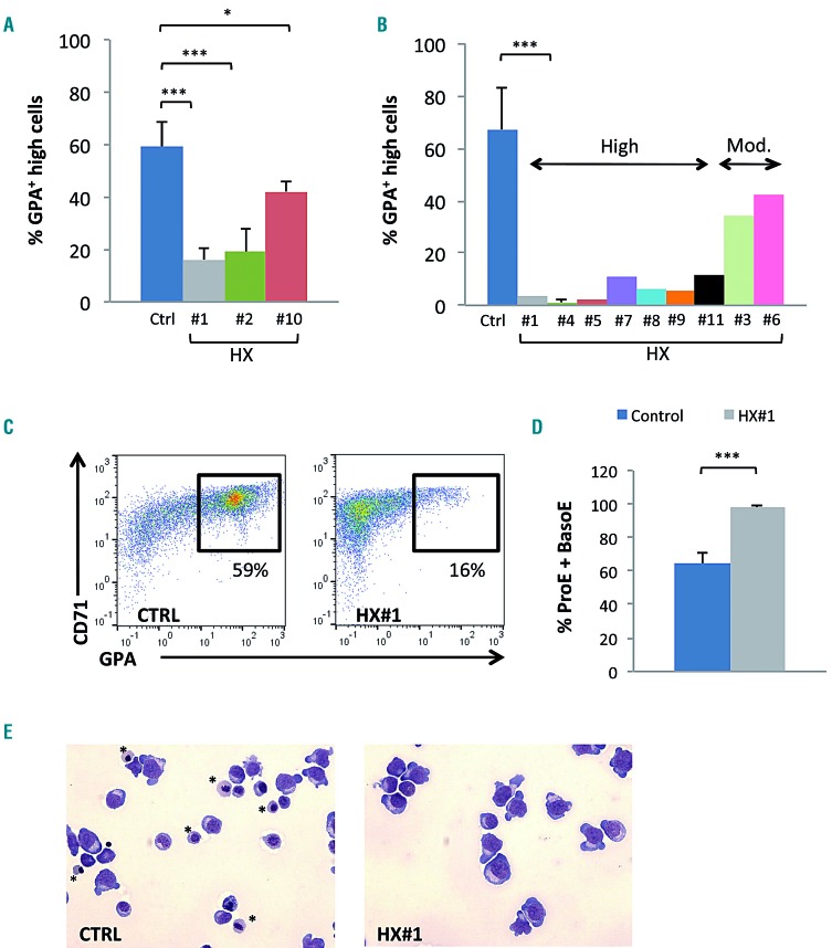
Delay in erythroid differentiation of progenitor cells obtained from patients with PIEZO1-mutated hereditary xerocytosis. (A) Culture of CD34+-derived erythroid cells from patients with hereditary xerocytosis (HX); differentiation was assessed at day 10 by multiparametric flow cytometry (MFC). The mean percentage of CD71+/GPAHigh cells was 59±9% in control samples, 16±5% in HX#1 (P<0.001), 19±9% in HX#2 (P<0.01), and 42±4% in HX#10 (P<0.05). (n=5 for control samples; n=3 for HX#1, 2 and 10). (B) Culture of mononuclear cells from HX patients; differentiation assessed on day 10 by MFC (n=9 for control samples; n=3 for HX#4; n=1 for HX#1-11). The mean percentage of CD71+/GPAHigh cells was 71±17% in the control group, and ranged from 0.3% to 11.7% in the “high delay” group and from 22.5% to 44% in the “moderate delay” group. (C) Illustrative CD71/GPA MFC plots at day 10 of erythroid differentiation of CD34+ cells obtained from patient HX#1 (right) and from one representative control sample (left). (D) Cytological analysis after staining with May-Grünwald-Giemsa (MGG): count of immature erythroblasts [proerythroblasts (ProE) and basophilic erythroblasts (BasoE)] on day 10, ×200 magnification. Immature erythroblasts were 64.2±6.2% in control samples vs. 97.7±1.5% in HX#1 (n=6 control samples; n=3 for patient HX#1). (E) Example of cytology on day 10 for patient HX#1 (right) and for a control sample (left), MGG staining, ×200 magnification. *show orthochromatic erythroblasts. ***P<0.001; **P<0.01; *P<0.05.

