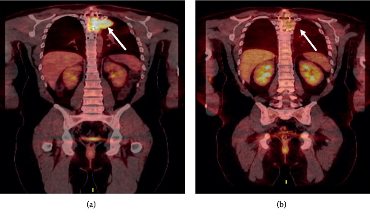
Figure 1.
(a) 18F FDG PET-CT at relapse showing large soft-tissue mass replacing the T4 vertebral body (white arrow). (b) 18F FDG PET-CT after 2 cycles of KRD-PACE showing near-complete resolution of the extraosseous soft-tissue component involving patient's known T4 vertebral body lesion (white arrow).

