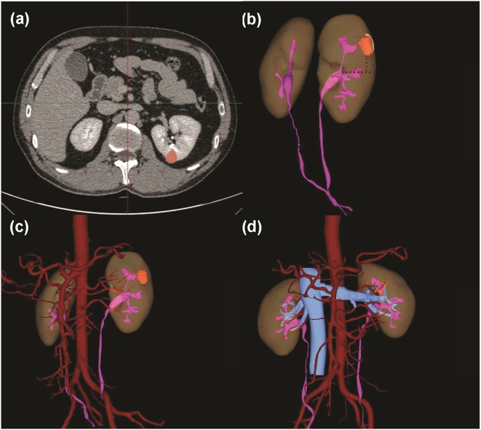
Figure 5. A case with a 3D score of 8.
(A) Tumor identification in 2D cross-section. (B) Total tumor volume: 2.05 ml; endophytic/exophytic volume rate: >50% endophytic; LDTE: 1.78 cm; tumor’s relationships with UCS or renal sinus: neither UCS nor sinus was involved; (C) Renal vascular variation: three feeding arteries of left kidney. (D) Relative position variation of renal artery and renal vein: no variation.

