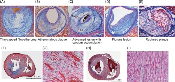
Fig. 3.

Photomicrographs of coronary lesions (A–E) and myocardial lesions (F–I) of WHHLMI rabbits
Panels A–D demonstrate spontaneously developed coronary lesions, and panel E demonstrated a ruptured coronary lesion after spasm provocation. Panels G and I are magnified photomicrographs of square in panel F and panel H, respectively. Panels A-F, and H show Azan staining, and panels G and H show HE staining. Panels A and E are modified from Shiomi M, et al.22), panels B–D are modified from Shiomi M, et al.23), and panels F-I are modified from Shiomi M, et al.4).
