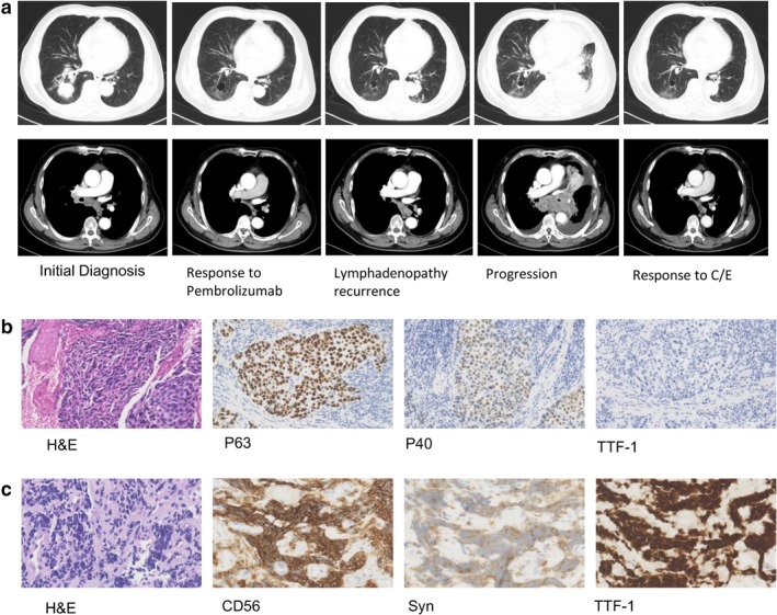
Figure 1.

(a) On initial diagnosis, chest computed tomography (CT) scan showed a 3.9 cm right lower lobe tumor with right hilar and mediastinal lymphadenopathy, and pericardial effusion. The patient was subsequently treated with pembrolizumab monotherapy and had a partial response. After 22 cycles of pembrolizumab, follow‐up CT scan showed a left hilar tumor, bilateral pleural effusion and lymphadenopathy recurrence. After two cycles of chemotherapy (carboplatin/etoposide/), CT scan revealed shrinkage of the lesions. (b) Pathologic findings from the right lower lobe lesion at the time of initial diagnosis showed squamous cell carcinoma (hematoxylin and eosin [H&E] staining, ×200), with positive immunohistochemical staining for P63 and P40 (×200), and negative staining for thyroid transcription factor1 (TTF‐1). (c) Pathologic finding from the intraluminal lesion in the left main bronchus at the time of progression showed small cell lung cancer (H&E, ×400), with positive immunohistochemical staining for CD56 (×400), synaptophysin (Syn×400), and TTF‐1(×400).
