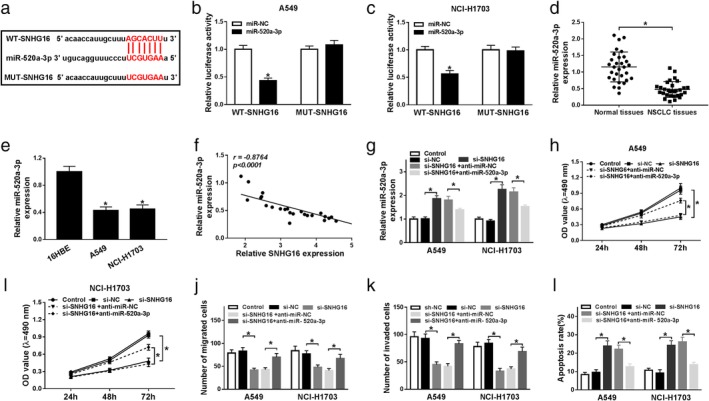
Figure 3.

SNHG16 silence exerted antitumor effects in NSCLC cells by directly sponging miR‐520a‐3p. (a) A graphical representation of the miR‐520a‐3p binding site within SNHG16 is presented. (b, c) The interaction between miR‐520a‐3p and SNHG16 was confirmed using a dual‐luciferase reporter assay in A549 and NCI‐H1703 cells. (d, e) The expression of miR‐520a‐3p was detected in normal tissues and NSCLC tissues (d), as well as NSCLC cell lines (A549 and NCI‐H1703) and human bronchial epithelial (16HBE) cell line (e) using qRT‐PCR. (f) The correlation between miR‐520a‐3p and SNHG16 expression was assessed in NSCLC tissues by Spearman's correlation test. A549 and NCI‐H1703 cells were transfected with si‐NC, si‐SNHG16, si‐SNHG16 + anti‐miR‐NC, or si‐SNHG16 + anti‐miR‐520a‐3p. After transfection, (g) the level of miR‐520a‐3p was measured in A549 and NCI‐H1703 cells by qRT‐PCR; (h, i) MTT assay was used to detect cell proliferation; (j, k) cell migration and invasion were determined by transwell assay; (l) apoptotic cells were examined by flow cytometry. *P < 0.05.
