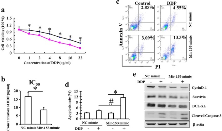
Figure 2.

miR‐153‐3p enhances the sensitivity to cisplatin. (a) Eca109 cells transfected with control or miR‐RNA‐153‐3p mimics were treated with different concentrations of cisplatin (0–32 μg/mL) for 24 hours, and the cell viability was measured by CCK8 assay. ( ) NC mimic, (
 ) Mir153 mimic. (b) IC50 of cisplatin in cells is shown. (c,d) Eca109 cells transfected with control or miR‐RNA‐153‐3p mimics were treated with 2 μg/mL cisplatin for 24 hours, and cell apoptosis was detected by FCM with double staining by Annexin and PI. Mean (± standard deviation) values from three independent experiments are shown (*P < 0.05 vs. control; #P < 0.05, control cells treated with cisplatin vs. microRNA‐153‐3p mimics cells treated with cisplatin). (e) Results of western blot showing the expressions of CyclinD1 and Survivin in cells.
