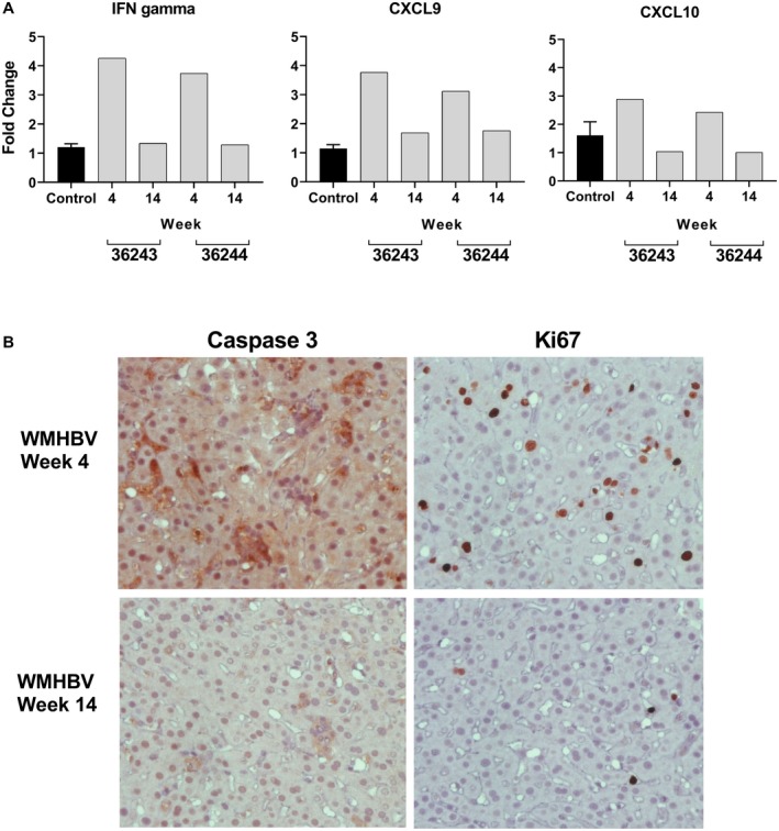
Figure 6.

Changes in IFN gene expression and markers of cell death and proliferation in the liver of AAV‐WMHBV‐infected monkeys. (A) TaqMan RT‐PCR was performed on total liver RNA from four control animals (C) and two animals infected with AAV‐WMHBV (36244, 36245) at 4 and 14 weeks following infection. Primers and probes for squirrel monkey IFN gamma, CXCL9 (i.e., Mig), and CXCL10 (i.e., IP‐10) were designed based on the genomic sequence of squirrel monkeys as described in the Materials and Methods section. The values for infected animals are expressed as a fold change in comparison to the controls. C is the average of four animals and is expressed as a fold change of 1, no fold change, with the range shown. (B) Immunohistochemical staining of liver sections from an AAV‐WMHBV‐infected monkey at week 4 and 14 for the cell death marker–activated caspase‐3 (left, upper, and lower panels) and for the cell proliferation maker Ki67 (right, upper, and lower panels).
