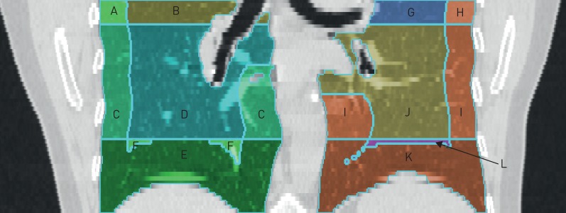
FIGURE 1.
Multi-Ethnic Study of Atherosclerosis (MESA) cardiac computed tomography scan with superimposed lines to delineate examined lung regions. In this representative scan, the basilar region includes sections E, F, K, L. The non-basilar region includes sections A, B, C, D, G, H, I, J. The peel includes sections A, C, E, H, I, K. The core includes sections B, D, F, G, J, L. The basilar peel includes sections E, K. The basilar core includes sections F, L.

