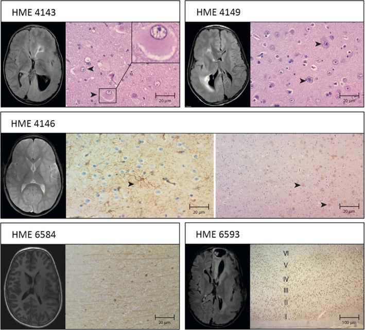
Figure 2.

Somatic‐positive HME cases exhibit MRI and histopathological findings. A, Tissue shows the presence of balloon cells by coloration hematoxylin and eosin and pre‐op FLAIR MRI left and right cerebral cerebellar volume increase, with lateral ventricular dilatation and periventricular hypersignal areas. B, Tissue shows cytomegalic and dysmorphic neurons, with accumulation of Nissel's substance at the periphery coloration hematoxylin/eosin, and pre‐op FLAIR MRI shows right hemisphere enlargement, cortical thickening, lateral ventricular dilatation, and periventricular hypersignal areas. C, Astrogliosis immunostained to GFAP and pre‐op T2 MRI shows evidence of signal change and morphology throughout the left hemisphere. D, Tissue shows the presence of heterotopic neurons in the white matter neocortex transition, immunostained to NeuN, and (E) tissues show neurofilaments accumulating in the body of dysplastic neurons immunostained for NeuN, and pre‐op FLAIR MRI shows increased right brain hemisphere volume and lateral ventricular dilatation. F, Tissue shows disorganization of the cortical layers IV, V, VI with neuronal loss by immunostaining for NeuN, and pre‐op FLAIR MRI shows increased left cerebral hemisphere volume, cortical thickening, lateral ventricular dilatation, and periventricular hypersignal areas
