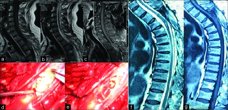
Figure 3:
(a) Cervicothoracic T1-weighted magnetic resonance imaging (MRI) the tumor is isointense, (b) in T2-weighted mage it is hypointense which is unusual for intramedullary epidermoid cysts, (c) but in fat-suppressed MRI the hyperintense mass is compatible with epidermoid, (d) intraoperative photograph shows the characteristic features of and epidermoid cyst, (e) after total removal, (f) T1-weighted MRI at 10-year follow-up shows neither residue nor recurrence, (g) T2-weighted image also is clear.

