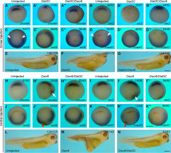
Figure 2.
DSCR6 functionally interacts with Stat3 in DV patterning. A–D″, in situ hybridization analysis shows the expression patterns of dorsal (chordin and otx2) and ventral (wnt8 and Xvent1) mesoderm genes in early gastrula stage embryos expressing Stat3C or Stat3C and DSCR6 in the dorsal region. Arrowheads indicate the dorsal limit of wnt8 expression in uninjected embryos. All embryos are vegetal view with dorsal region up. E–G, live images at larval stage (stage 36) show that DSCR6 rescues anterior deficiency produced by dorsal activation of Stat3C. H–K″, in situ hybridization analysis shows the expression patterns of dorsal and ventral markers in early gastrula stage embryos expressing DSCR6 or DSCR6 and Stat3C in the ventral region. Arrows indicate ectopic expression of chordin and otx2; arrowheads show the repression of wnt8 and Xvent1 expression. All embryos are vegetal view with dorsal region up. L–N, live images at stage 36 show that DSCR6-induced formation of secondary axis is blocked by activation of Stat3. Scale bars: A–D″) 500 μm; E–G, 500 μm; H–K″, 500 μm; L–N, 500 μm.

