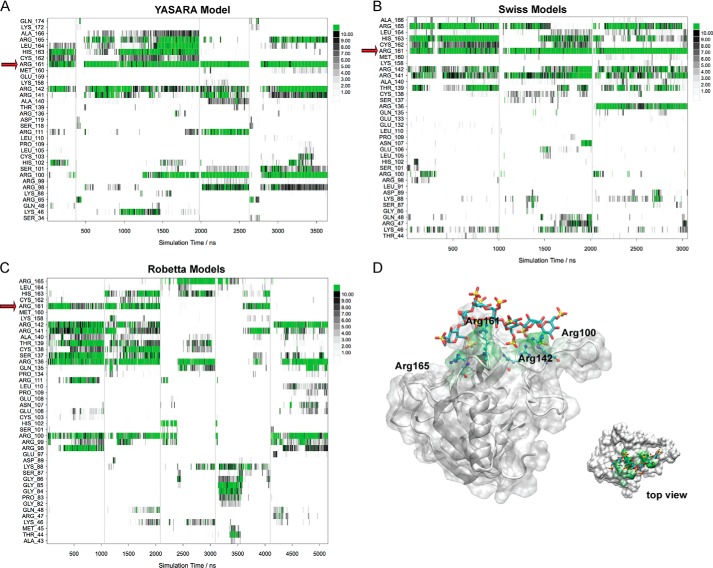
Figure 8.
Molecular dynamics simulations of the models of the lectin domain of CLEC14A. A–C, accumulated trajectory plots of the number of heavy atom contacts (<4.0 Å) between the heparin fragment and the individual amino acids of CLEC14A. A vertical line is used to separate individual simulations. Amino acids that did not participate in any intermolecular interactions in the simulations are not shown in the plots. Red arrows indicate Arg-161. A, YASARA models; B, Swiss models; C, Robetta models. D, computational prediction of the heparin-binding site of the lectin domain of CLEC14A (YASARA model). Amino acids that participated in strong interactions with the heparin fragment during the MD simulations (accumulated 3.6 μs) are highlighted in green.

