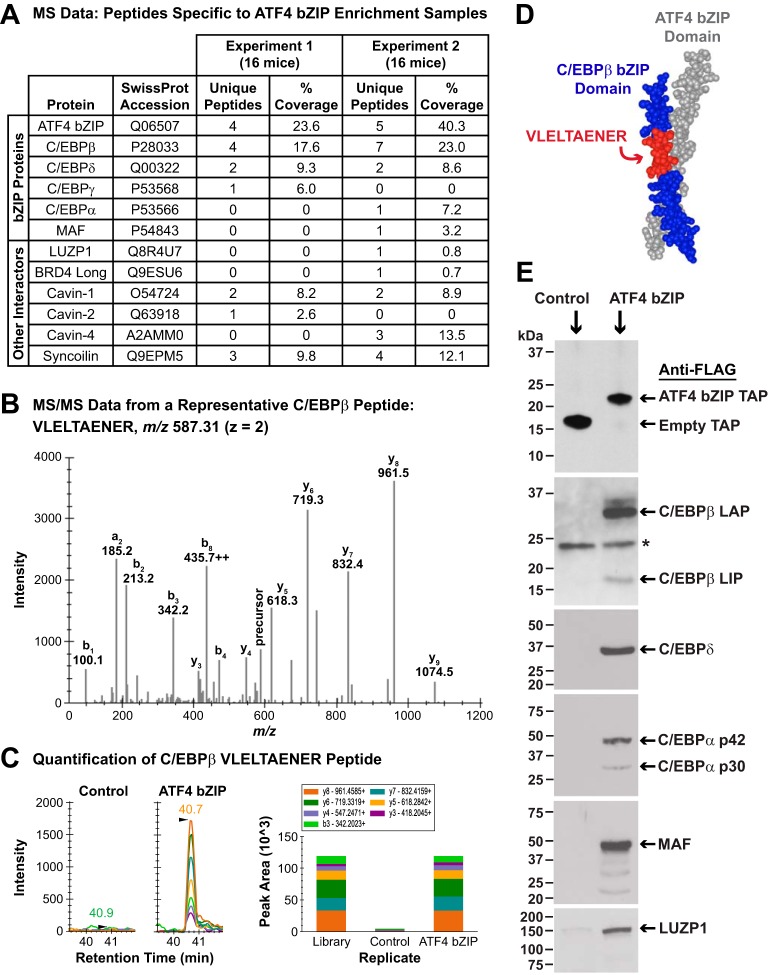
Figure 2.
Identification of proteins that interact with the ATF4 bZIP domain in mouse skeletal muscle fibers. A, control and ATF4 bZIP pulldown samples from two independent, time-separated experiments were obtained as described in Fig. 1D and then subjected to MS and a data analysis workflow that is fully detailed under “Experimental procedures” and the supporting information. The table in A summarizes the mass spectrometric data for ATF4 and the high-confidence ATF4-interacting proteins that were identified in the ATF4 bZIP pulldown samples. For proteins with only one peptide identified, the corresponding MS/MS were manually inspected (also see under “Experimental procedures” and Fig. S2). B, tandem mass spectrum (MS/MS) from a representative C/EBPβ peptide VLELTAENER (SwissProt accession P28033) spanning residues 254–263 with the precursor ion at m/z 587.31 2+ identified in the ATF4 bZIP affinity-enrichment sample. C, left, quantification of C/EBPβ peptide VLELTAENER using DIA MS2-based quantification; panels show extracted ion chromatograms for the MS2 fragment ions in the control and ATF4 bZIP affinity enrichment sample. Right, MS/MS spectral library “simulation” indicating relative fragment ion distribution and calculated peak areas under the curve for the C/EBPβ peptide VLELTAENER in control and ATF4 bZIP affinity enrichment samples. D, mapping of VLELTAENER to the crystal structure of the ATF4 and C/EBPβ bZIP domains (19). E, TA muscle fibers of 12 mice were transfected with 20 μg of empty TAP plasmid (one TA per mouse) or 20 μg of ATF4 bZIP TAP plasmid (the contralateral TA in each mouse). Seven days post-transfection, bilateral TA muscles were harvested and used to prepare pooled protein extracts from each of the two groups of skeletal muscles (control and ATF4 bZIP). The pooled protein extracts were then subjected to pulldown with anti-FLAG magnetic beads, followed by SDS-PAGE and immunoblot analysis using HRP-conjugated mouse monoclonal anti-FLAG IgG (upper panel), mouse monoclonal anti-C/EBPβ IgG, rabbit polyclonal anti-C/EBPδ antibody, rabbit monoclonal anti-C/EBPα IgG, rabbit polyclonal anti-MAF antibody, and rabbit polyclonal anti-LUZP1 antibody, as indicated. Asterisk in the anti-C/EBPβ immunoblot denotes a nonspecific cross-reacting protein.

