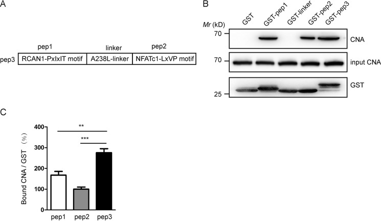
Figure 1.
The binding capacity between CNA and pep3 is more potent in mouse brain lysates. A, schematic representation of pep1, pep2, linker, and pep3. B, pep3 binds CNA in mouse brain lysates in GST pulldown assays. Bound CNA was visualized by Western blotting via monoclonal anti-CNA antibody, shown in the upper blot; the input is shown in the middle panel. The GST fusion proteins were shown in the bottom blot. C, CNA bound by pep1, pep2, and pep3 was densitometrically quantified, and histograms illustrate the relative intensity units of bound CN. Data are presented as the mean ± S.E. (n = 3), **, p < 0.01; ***, p < 0.001 compared with the pep3 group.

