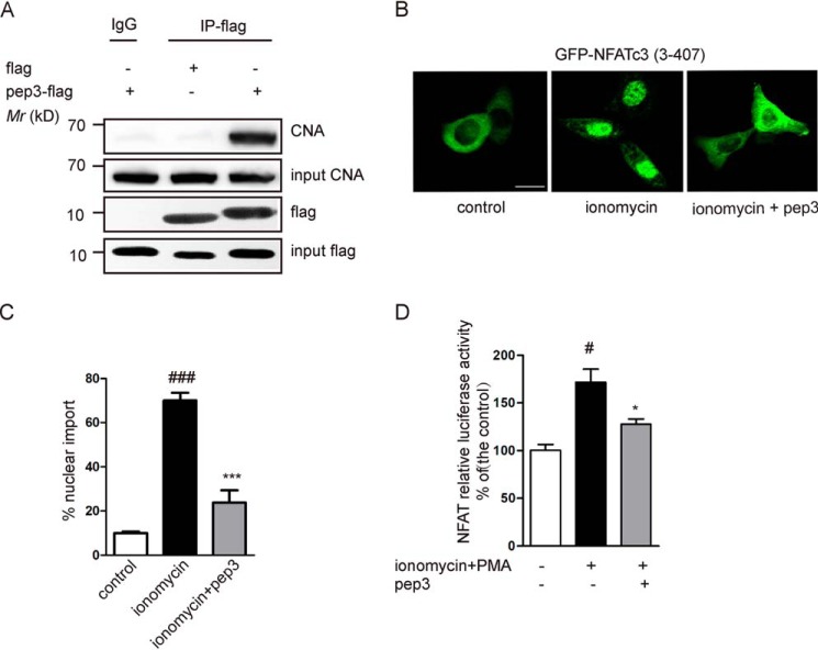
Figure 3.
Expression of pep3 suppresses CN-NFAT signaling pathway. A, Western blotting was utilized to detect CNA pulled down by an anti-DDK (flag) antibody in pep3-flag–transfected HEK293 cells. B, GFP-NFATc3 (3–407) plasmids were transfected into HeLa cells. Representative images shows that transfection of pep3-flag blocks ionomycin-induced GFP-NFATc3 translocation. Scale bar, 10 μm. C, bar graph shows the percentages of NFAT nuclear translocation in HeLa cells. Data were presented as mean ± S.E. of four independent experiments with n = 100 cells per experiment, ###, p < 0.001 compared with the control group; ***, p < 0.001 compared with the ionomycin group. D, pep3 inhibits NFAT-driven gene expression. HEK293 cells were transiently transfected with pep3-flag, together with pGL3-NFAT luciferase. pRL-null-Renilla-luc (7.5 ng) was used as an internal transfection control. Cells were stimulated with ionomycin + PMA for 1.5 h. Control values were taken as 100%. All data are expressed as the mean ± S.E. of three separate experiments. #, p < 0.001 compared with the control group; *, p < 0.05 compared with the ionomycin + PMA group.

