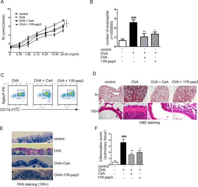
Figure 5.
Intranasal administration of 11R-pep3 inhibits airway inflammation and hyperresponsiveness. A, airway reactivity was assessed by MCh provocation testing. The mean ± S.E. is presented for each group (n = 6). *, p < 0.05 compared with the OVA group. B, total numbers of eosinophils in leukocytes of BAL fluids. Bar graph depicts the numbers of eosinophils in leukocytes of BAL fluids for each group. Data are presented as the mean ± S.E. for each group (n = 6). ###, p < 0.001 compared with the control group; *, p < 0.05; **, p < 0.01 compared with the OVA group. C, representative results of flow cytometry methods. We defined eosinophils in leukocytes as CD45+CD11b+SiglecF+. D, lung tissue was prepared for histological analysis, including morphology and the infiltration of inflammatory cells by H&E staining. Representative results of H&E staining in four groups are shown. Scale bars, 200 μm (top) and 10 μm (bottom). E, lung tissue was prepared for histological analysis. PAS-stained airway cross-sections of each group are shown. Scale bar, 10 μm. F, inflammatory changes were graded by histopathological assessment using a semiquantitative scale of 0–5. The results are presented as the mean ± S.E. for each group (n = 6).

