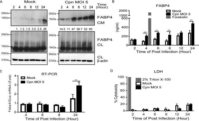
Figure 1.
C. pneumoniae infection induces the secretion of FABP4 from murine adipocytes. A, immunoblot analysis of FABP4 in cultured medium (CM) and cell lysates (CL) of 3T3-L1 adipocytes after mock or C. pneumoniae (Cpn) infection for 2–24 h. β-Actin served as the standard. B, secretion of FABP4 was measured in the cultured medium of 3T3-L1 adipocytes after mock or Cpn infection for 2–24 h. A 4-h incubation with forskolin (20 μm) served as the positive control for lipolysis. C, relative levels of Fabp4 mRNA in 3T3-L1 adipocytes after mock or Cpn infection for 4–24 h, as determined by real-time PCR. Gus mRNA served as the internal control. D, LDH assay using the supernatant of 3T3-L1 adipocytes at 2–24 h after mock or Cpn infection (n = 3/group; B–D). **, p < 0.01 by two-way ANOVA (B and C). The data are shown as the means ± S.E. and are representative of at least three experiments.

