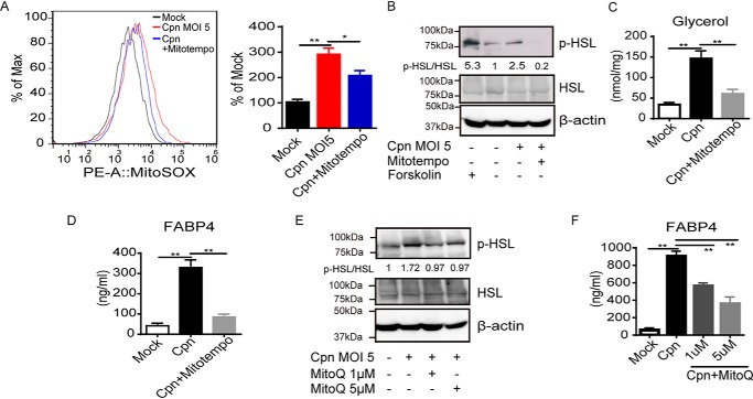
Figure 3.
C. pneumoniae infection–induced FABP4 secretion depends on mitochondrial ROS. A, flow cytometry (left panel) and quantification (right panel) of MitoSOX-stained 3T3-L1 adipocytes at 24 h after Cpn infection at a MOI of 5 in the presence or absence of MitoTEMPO (100 μm). B and E, immunoblot analysis of p-HSL and HSL in cell lysates of 3T3-L1 adipocytes at 24 h after Cpn infection in the presence or absence of MitoTEMPO (100 μm) (B) or increasing doses of MitoQ (E). A 2-h incubation with forskolin (20 μm) served as the positive control for lipolysis. β-Actin served as the standard. C, glycerol levels were measured in cultured medium of 3T3-L1 adipocytes at 24 h after Cpn infection in the presence or absence of MitoTEMPO. D and F, FABP4 levels in cultured medium of 3T3-L1 adipocytes at 24 h after Cpn infection at a MOI of 5 in the presence or absence of MitoTEMPO (100 μm) (D), increasing doses of MitoQ (Mitoquinone) (F). In A, C, D, and F, n = 3/group. *, p < 0.05; **, p < 0.01, one-way ANOVA (A, C, D, and F). The data are shown as the means ± S.E. and are representative of at least three experiments.

