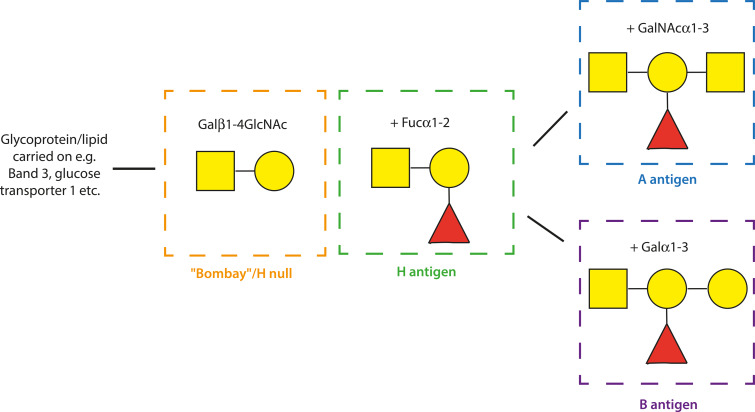
Fig. 3.
Diagram of the ABO blood group sugars. Schematic representation of the terminal structure of the A (blue square), B (purple) H (green; H is the antigen carried on blood group O erythrocytes) and Bombay (orange) antigens. Yellow circle: D-Galactose (Gal), yellow square: N-acetyl-D-galactosamine (GalNac), red triangle: L-Fucose (Fuc). The symbols α and β indicate the position of the hydroxyl group and the numbers indicate the specific carbon atoms that are linked between the sugars. The H, A and B antigens are synthesized by a series of glycosyltransferase enzymes that add monosaccharides to create oligosaccharide chains attached to lipids and proteins in the erythrocyte membrane.

