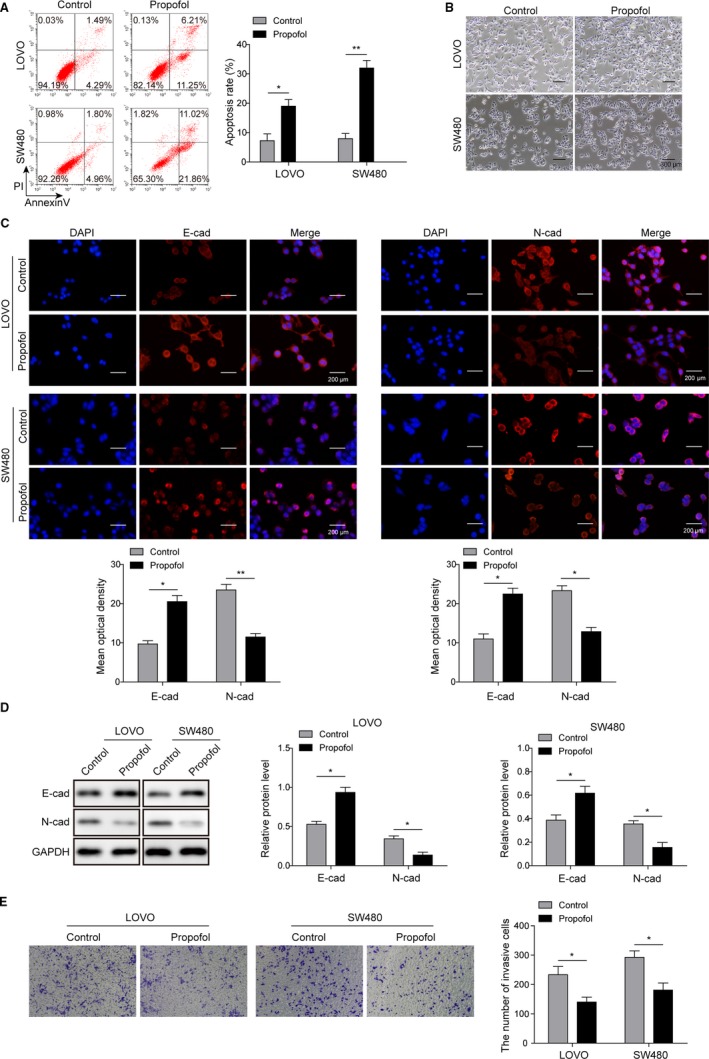
Figure 1.

Propofol induced the apoptosis of colon cancer cell lines and inhibited their invasion. A, Flow cytometry showed that apoptotic rate of both LOVO and SW480 were significantly increased after propofol treatment; *P < .01 and **P < .05 vs Control. B, Propofol‐treated LOVO and SW480 cells showed less mesenchymal and more epithelial phenotype compared to control; Scale bar, 300 µm. C, Immunofluorescence staining was subjected to detect the expression of E‐cadherin and N‐cadherin in both LOVO and SW480 cells treated with propofol; Scale bar, 200 µm. *P < .01 and **P < .05 vs Control. D, Western blot analysis was performed to determine the expression of E‐cadherin and N‐cadherin in both LOVO and SW480 cells treated with propofol. *P < .05 vs Control. E, Transwell assay showed decreased invasive cell number after propofol treatment in both LOVO and SW480 cells. *P < .05 vs Control
