Fig. 5.
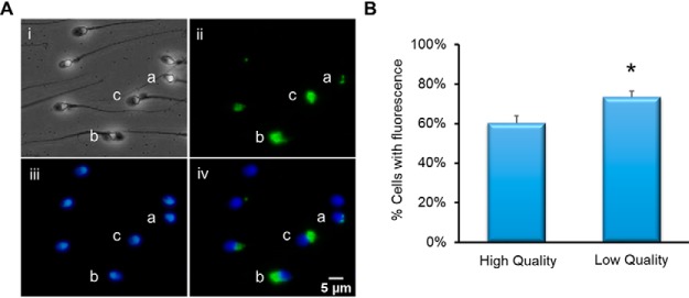
Immunocytochemical analysis of PDIA3. Spermatozoa were fixed and settled onto poly-lysine slides. Following permeabilization with 0.2% Triton-X and blocking with 3% BSA, the cells were incubated with primary anti-PDIA3 antibody then secondary alexafluor-488-conjugated antibody and counterstained with DAPI. A, (i) Phase image of spermatozoa, (ii) Immunofluorescent image using anti-PDIA3 antibody. Staining can be observed in a- redundant nuclear envelope, b-perinuclear theca, c-residual cytoplasm. (iii) DAPI staining of spermatozoon DNA, (iv) composite image of DAPI and PDIA3 fluorescence. Scale bar = 5 μm. B, Comparison of cells with detected PDIA3 fluorescence between high- and low-quality cell populations (n = 3, *p = <0.01).

