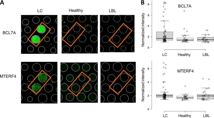
Fig. 2.
Examples of IgA-bound autoantigens identified on HuProt arrays in Phase I. A, Anti-human IgA images of BCL7A and MTERF4 obtained with serum samples collected from a LC patient, healthy subject, and LBL patient. IgA-bound autoantibodies were visualized with a Cy3-labeled anti-human IgA secondary antibody on HuProt arrays. In both cases, BCL7A and MTERF4 were specifically recognized by IgA antibodies of a LC patient; no detectable signals were observed with a healthy or LBL serum. B, Box plot analysis of HuProt array profiling of BCL7A (upper panel) and MTERF4 (lower panel) in LC, healthy and LBL.

