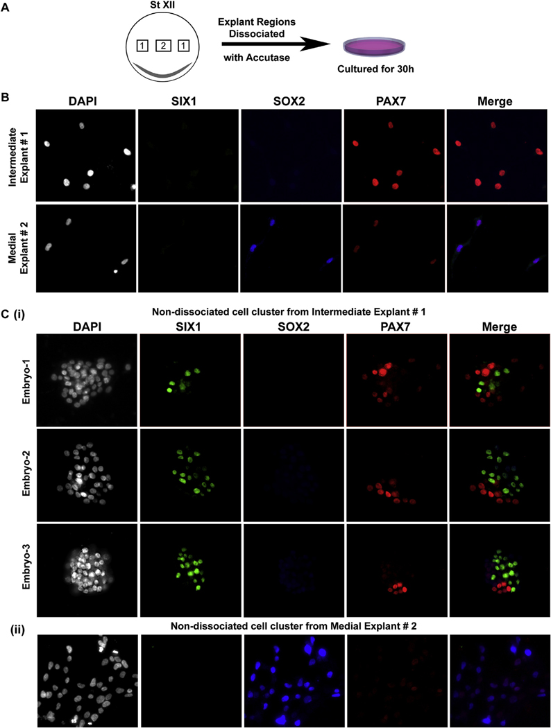
Fig. 4. Intermediate epiblast consists of precursors of NC and placodal cell fates.
(A) Schematic showing intermediate and medial explant regions from EGK stage XII used for the analysis. (B) Explants from EGK St. XII epiblast were dissociated and cultured for 36hrs. Distant cells in this low-density culture from intermediate explant (explant #1) express Pax7, no ectodermal marker was expressed in these cells. Distant cells in low density culture from medial explants (explant #2) expressed Sox2 and lower levels of Pax7. Expression of placodal gene, Six1, in distant isolated cells was not seen. (C) (i) Images from 3 individual embryos, from few remaining undissociated clusters of cells in the culture corresponding to intermediate epiblast region, analyzed for Six1, Sox2 and Pax7 expression. Six1 is expressed in cells present in clusters. Six1 and Pax7 expression is mutually exclusive. (ii) Undissociated clusters of cells in the culture corresponding to medial epiblast region, display robust Sox2 expression and weak Pax7 expression. This expression pattern was observed in dissociated intermediate explants from 6 individual embryos. Cell counts for Six1+ cells are provided in supplemental Table 1.

