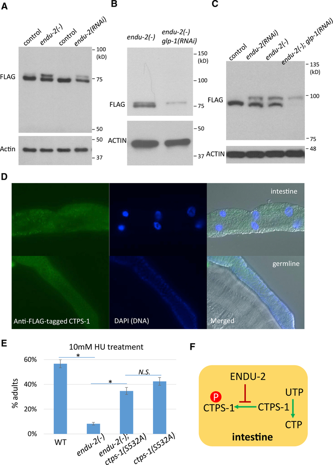
Figure 3. ENDU-2 Negatively Regulates CTPS-1 Phosphorylation in the Intestine.
(A) Western blot of FLAG::CTPS-1 in samples from indicated strains. Two bands were observed in both endu-2(−) and endu-2(RNAi) worms. The flag::ctps-1 background is included in all of the experiments for the detection of CTPS-1 (see Method Details). Samples were run on a 7.5% SDS-PAGE gel.
(B and C) Western blot of FLAG::CTPS-1 in endu-2(−) mutant worms treated with control or glp-1 RNAi that eliminates the germline. Samples were run on a 12% SDS-PAGE gel (B) or a 12.5% phos-tag gel (C) (see Method Details).
(D) Immunostaining of FLAG::CTPS-1 showed the expression of a FLAG-tagged CTPS-1 in the intestine (top panels) and the germline (bottom panels). See Figure S3A for negative controls.
(E) Disrupting the predicted phosphorylation site of CTPS-1 significantly suppressed the hypersensitivity of endu-2(−) animals to HU treatment. Error bars indicate standard deviation. Asterisks indicate significant differences, N.S., not significant. Tukey’s range test, FWER = 0.01.
(F) A model based on the genetic interaction and biochemical assays that show that ENDU-2 positively regulates CTPS-1 by blocking its phosphorylation.

