
Figure 1.
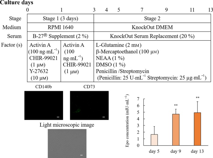
Erythropoietin concentration in the supernatant and overview of the modified differentiation protocol. The differentiation protocol was performed by modifying a previous method 9. hiPSCs grown on plates coated with iMatrix‐511 were cultured in StemFit medium for 7 days. The hiPSCs were dissociated to single cells by gentle pipetting after treatment with Accutase and seeded on Matrigel‐coated plates with stage 1 medium containing RPMI 1640 supplemented with B27 supplement, recombinant human/mouse/rat activin A, and 1 μm CHIR99021. After 24 h, this medium was replaced with fresh medium without Y‐27632 until culture day 3. The medium was changed to stage 2 medium containing KnockOut DMEM supplemented with penicillin/streptomycin, 20% KSR, 1% DMSO, 2 mm l‐glutamine, 1% NEAA, and 100 μm β‐mercaptoethanol. The hiPSCs were stained with anti‐CD140b (red color) and anti‐CD73 (green color) antibodies. The white bar indicates 100 µm. EPO concentrations are shown as mean values ± SD. **P < 0.01 vs. day 5 (Bonferroni's test).