
Figure 5.
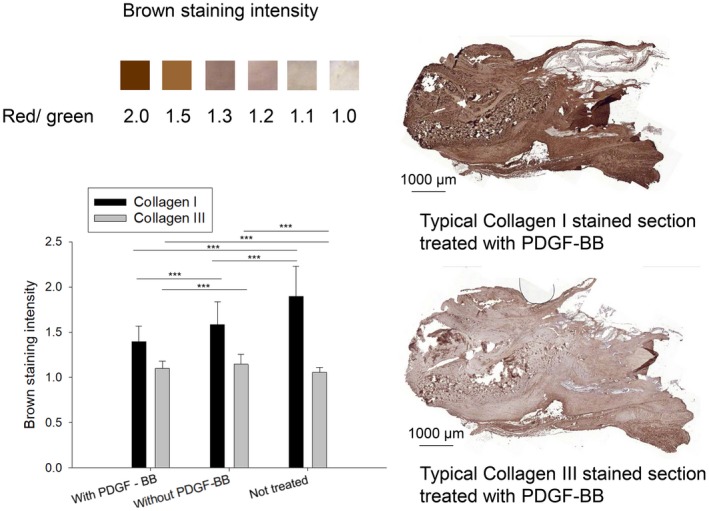
Collagen I and III immunohistochemistry of Achilles tendons in paraffin sections, with red/green ratio to assess brown staining intensity and typical examples for tendons stained with collagens I and III. One‐way ANOVA was performed. P values < 0.05 were considered significant (*). If P < 0.01, this was marked by **, and if P < 0.001 by ***. Error bars indicate standard deviations. Biological independent replicates n = 6. Scale bars = 1000 µm. Positive (NT) and negative (rabbit brain) controls for collagen I and III stainings are given in Fig. S6.