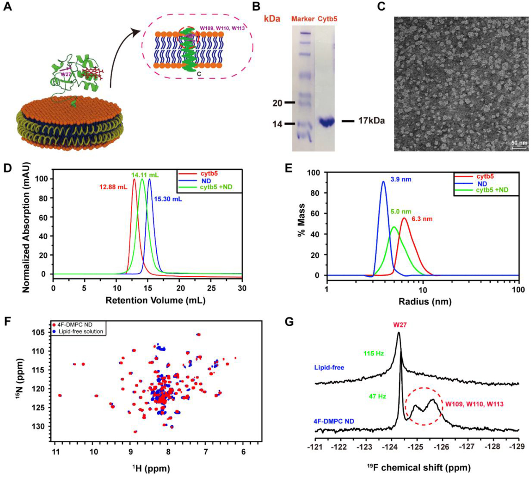
Figure 1. Reconstitution of cytochrome b5 in peptide-based lipid-nanodiscs.

(A) Ribbon diagrams of cytochrome b5 in a nanodisc indicating 5F-tryptophan-labeled sites. (B) SDS-PAGE gel showing the purity 5FW-labeled cytb5. (C) TEM image of 4F-DMPC nanodiscs with a diameter of ~10 nm. (D) SEC elution profiles and (E) DLS profiles of cytb5 alone in buffer (red), empty 4F-DMPC nanodiscs (blue), and 4F-DMPC nanodiscs containing cytb5 (green). (F) 2D 1H-15N HSQC spectra of 15N-labeled cytb5 alone in buffer (blue) and reconstituted in the 4F-DMPC nanodiscs (red). (G) 1D 19F NMR spectra of 5F-tryptophan-labeled cytb5 alone in buffer (top trace) and reconstituted in the 4F-DMPC nanodiscs (bottom trace) at 298K.
