Figure 3.
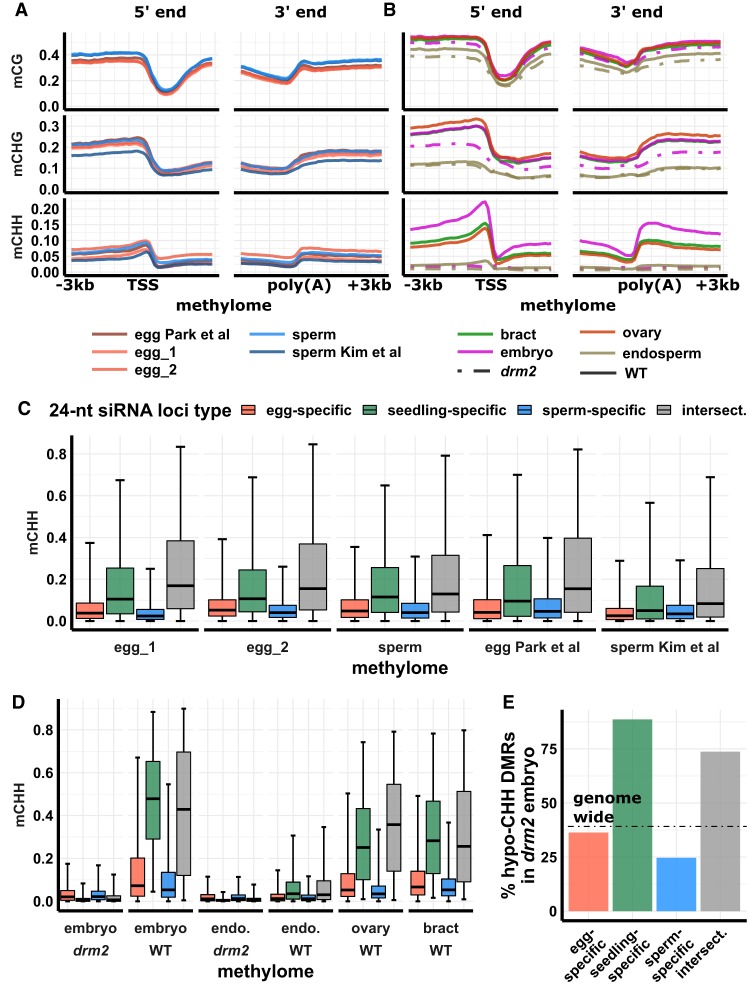
DNA methylation across tissues. (A) DNA methylation metagene plots for post-bisulfite adapter tagging (PBAT) libraries. Plots indicate average DNA methylation values over 100-bp intervals. Vertical grid lines indicate 500-bp intervals. DNA methylation is measured as the proportion of methylated cytosines relative to total cytosines in each sequence context. (TSS) Transcription start site, (poly[A]) polyadenylation site. Published data from Park et al. (2016) and Kim et al. (2019). (B) DNA methylation metagene plot for conventional WGBS libraries; x- and y-axes as in A. (C) DNA methylation of 24-nt siRNA loci for PBAT libraries. Center line is median; box spans interquartile range; whiskers span 2.5th to 97.5th percentiles. (D) DNA methylation of 24-nt siRNA loci for conventional WGBS libraries, as in C; color code as in C. (E) Abundance of hypomethylated CHH differential methylated regions (hypo-CHH DMRs) in drm2 mutant relative to wild-type mature embryo. Abundance is measured as the percent of hypomethylated regions relative to total number of regions with sufficient read coverage for statistical significance. Dotted line indicates genome-wide average percent hypo-CHH DMRs. Color code as in C.

