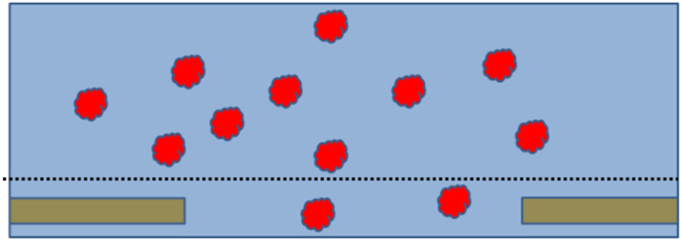
Figure 1.
Cartoon of what an ideal cryo-EM specimen might be like. Depicted here is just a single hole in a thin, holey-carbon support film, which itself is supported by an EM grid (not shown). An excess amount of sample is first applied to the carbon film, after which everything above an imaginary dotted line is blotted away with filter paper. Before blotting, the biological macromolecules – represented by the red particles – are distributed randomly in suspension, and their distribution within the remaining sample, after blotting, is not imagined to be affected by removal of material above the dotted line. After freezing, the particles are embedded in vitreous ice, thus providing a sample whose structure is nearly identical to what it was in bulk solution. This is what had long been thought to be true, but we now know that it frequently is not what really happens.

