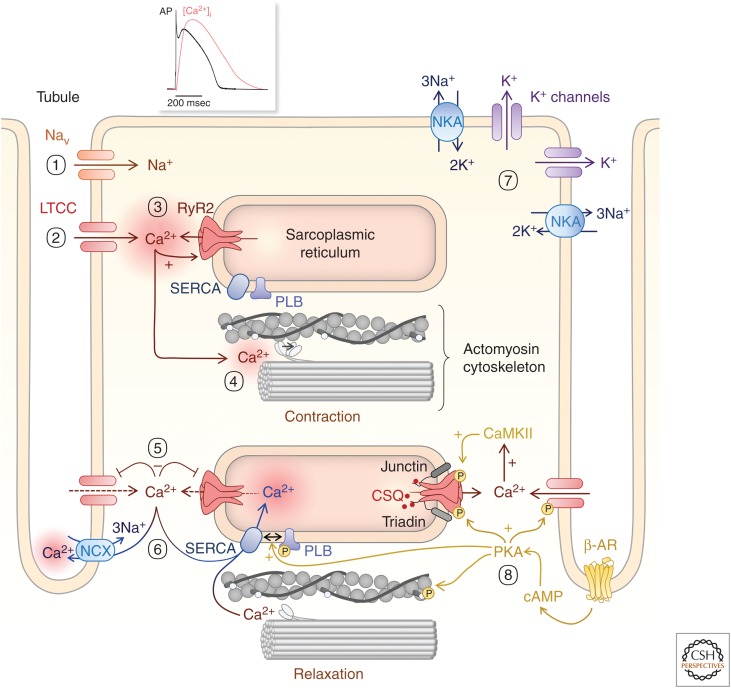
Figure 1.
Excitation–contraction coupling (ECC). (1) An action potential depolarizes the cardiomyocyte and induces Na+ influx through voltage-gated Na+ channels (Nav). (2) This further depolarizes the cell membrane and induces Ca2+ influx through voltage-gated L-type Ca2+ channels (LTCCs). (3) This Ca2+ entry stimulates Ca2+ release via dyadic RyR2 on the SR (4), which in turn triggers cell contraction through activating myofilament crossbridges. (5) LTCC inactivate and RyR close. (6) Cytosolic Ca2+ is then moved out of the cell by the Na+/Ca2+ exchanger (NCX) and pumped back into the SR by SERCA2a, thereby decreasing cytosolic Ca2+ concentration and bringing about relaxation. (7) A family of K+ channels participate in cell repolarization with K+ efflux as a last step for returning membrane potential to its resting value before a new cycle starts. (8) Tuning ECC to meet cardiovascular demands involves β-adrenergic pathways that induce the activation of CaMKII and of cAMP/PKA, which phosphorylates voltage-gated LTCCs and RyRs to enhance their activity and phospholamban (PLB) to remove its inhibition of SERCA activity.

